The threshold potential (critical value of depolarization that can provoke an action potential) also becomes progressively less negative with a rising extracellular K concentration. As the external K increases, the ratio will decrease causing partial depolarization of the cellular membrane to a less electronegative value ( Figure 2). According to the Nernst equation or its simplified equivalent: 61 log K I/ K o (with I=intracellular and o=extracellular) the resting membrane potential or ionic equilibrium is directly related to the ratio of intracellular to extracellular K concentration. The potassium concentration gradient between extracellular and intracellular sites is the most important factor that controls the resting membrane potential. (Reproduced from Barold SS 30 with permission.) Electrophysiology 7 Convex ST elevation in the inferior leads during ventricular pacing has not been previously reported.

Such ST elevation may occur in hyperkalaemia usually involving the right precordial leads and less commonly in the inferior leads. There was no evidence of acute myocardial infarction and angiography revealed normal coronary arteries. Note the ST elevation in the inferior leads simulating an acute inferior myocardial infarction.

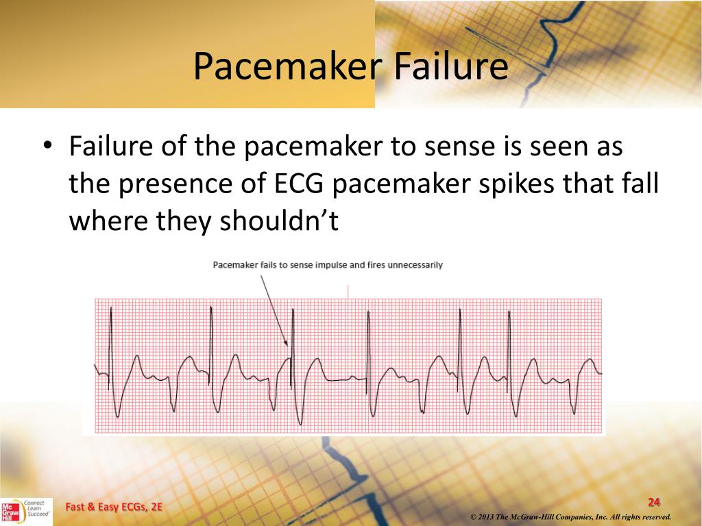
Some of the leads show pseudolatency of ventricular stimulation because of the initial isoelectric activation. Twelve-lead ECG during hyperkalaemia ( K = 7.2 mEq/L) showing loss of atrial capture and marked increase in the duration of the paced QRS complex (0.34–0.36 s). Failure to capture is usually seen when the K level reaches 7 mEq/L and occasionally at a lower level especially in the presence of heart disease. (ii) Increased atrial and ventricular pacing thresholds with or without increased latency. Other common causes of a wide paced QRS complex include amiodarone therapy and severe myocardial disease. When the K level exceeds 7 mEq/L, the intraventricular conduction velocity is usually decreased and the paced QRS complex widens. In patients with pacemakers, hyperkalaemia causes two important clinical abnormalities: (i) widening of the paced QRS complex (and paced P-wave if it is seen) on the basis of delayed myocardial conduction ( Figure 1). 1–6 Hyperkalaemia is the most common electrolyte or drug abnormality to cause loss of capture by a cardiac rhythm device. It is common in older patients undergoing intensive treatment for heart failure (with potassium-retaining drugs) especially if there is coexisting renal insufficiency. Hyperkalaemia may affect ∼8% of hospitalized patients in the USA. Hyperkalaemia, Cardiac pacemaker, Implantable cardioverter-defibrillator, Pacemaker failure, Oversensing by ICD A raised impedance from the right ventricular coil to the superior vena cava coil may become an important sign of hyperkalaemia in the asymptomatic or the minimally symptomatic ICD patient. The latter may cause inappropriate shocks. With implantable cardioverter-defibrillators (ICDs) oversensing of the paced or spontaneous T-wave may occur. During managed ventricular pacing, hyperkalaemia-induced marked first-degree atrioventricular block may induce a pacemaker syndrome. Ventricular undersensing is uncommonly observed because of frequent antibradycardia pacing. The disturbance may then progress to 2 : 1, 3 : 1 pacemaker exit block, etc., and eventually to complete exit block with total lack of capture. First-degree ventricular pacemaker exit block may progress to second-degree Wenckebach (type I) exit block characterized by gradual prolongation of the interval from the pacemaker stimulus to the onset of the paced QRS complex ultimately resulting in an ineffectual stimulus. In this respect, the atria are more susceptible to loss of capture than the ventricles, and (iii) Increased latency (usually with ventricular pacing) manifested by a greater delay of the interval from the pacemaker stimulus to the onset of depolarization.

In patients with pacemakers, hyperkalaemia causes three important abnormalities that usually become manifest when the K level exceeds 7 mEq/L: (i) widening of the paced QRS complex from delayed intraventricular conduction velocity, (ii) Increased atrial and ventricular pacing thresholds that may cause failure to capture.


 0 kommentar(er)
